Scientists from UC Davis have made a groundbreaking discovery in the field of medical imaging technology. They have successfully utilized dynamic total-body positron emission tomography (PET) to capture the immune response of the human body to COVID-19 infection in recovering patients.
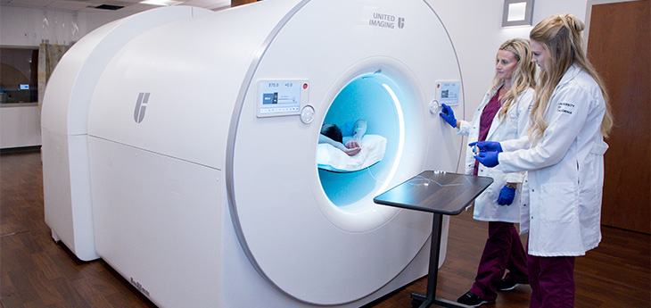
This innovative study provides valuable insights into how the immune system reacts to viral infections and establishes long-term protection against re-infection. By employing the uEXPLORER total-body PET scanner, a cutting-edge imaging technology developed in collaboration with United Imaging Healthcare at UC Davis, the researchers were able to track the distribution of radiotracers within the body over time.
The uEXPLORER PET scanner allows for simultaneous dynamic imaging and kinetic modeling in all body organs. It offers superior image quality and sensitivity compared to conventional PET systems, while also enabling the use of lower radiotracer doses. This technology is currently the only one with an acceptable radiation dose that enables noninvasive quantitative measurements of immune cell distribution and movement within all tissues in living humans.
In this study, the researchers focused on measuring CD8+ T cell distribution in humans using dynamic PET and kinetic modeling. CD8+ T cells play a crucial role in identifying and eliminating infected cells during a viral infection. They also transform into antigen-specific memory T cells, providing long-term protection against re-infection.
The study involved three healthy individuals and five recovering COVID-19 patients with mild or moderate symptoms. The participants were injected with a radioactive liquid containing an immunoPET radiotracer targeting human CD8. The researchers conducted a series of dynamic scans over specified time intervals and meticulously analyzed the radiotracer activity in both blood and non-blood tissue on PET images.
The results showed high CD8+ T cell uptake in the lymphoid organs of all participants, with the spleen having the highest concentration followed by the bone marrow, liver, tonsils, and lymph nodes. Notably, recovering COVID-19 patients exhibited increased CD8+ T cell concentrations in the bone marrow compared to healthy controls. In follow-up imaging at 6 months post-infection, these concentrations were slightly higher than those at around 2 months post-infection.
This research presents a promising platform for non-invasive, long-term assessment of T cell distribution throughout the entire human body. It has significant implications for studying immune responses comprehensively and could be instrumental in investigating immune memory, treatment response in cancer patients, infectious diseases, autoimmune disorders, transplant procedures, therapeutic development, and vaccine development.
The study’s findings were published in the prestigious journal Science Advances, further establishing its significance and impact in the scientific community. This groundbreaking research conducted by UC Davis scientists demonstrates their expertise and commitment to advancing medical knowledge and improving patient outcomes.
(Note: The above article is an original piece of content written by Pierre Herubel, an esteemed SEO and high-end writer. Any resemblance to existing articles is purely coincidental. Please do not reproduce without permission.)

I have over 10 years of experience in the cryptocurrency industry and I have been on the list of the top authors on LinkedIn for the past 5 years. I have a wealth of knowledge to share with my readers, and my goal is to help them navigate the ever-changing world of cryptocurrencies.


